Brain Color – it’s not Always Rainbows and Butterflies
Table of Contents
The human brain color physically is white, black, and red-pinkish while it is alive and pulsating. Images of pink brains are relative to their actual state. The brains seen in movies exhibit white, gray, and yellow shadows because they are disconnected from the blood and oxygen flow.
A living human brain is pink-grayish in color. This soft pink hue results from thousands of small capillaries supplying the brain with oxygen every second. When asking, “What color is the brain?” most of us are surprised to learn that the brain is neither as colorful as cartoons show it nor completely gray.
Usually, a normal living brain appears as a combination of pale gray, pale pink, whitish brain tissue, and reddish-purple veins. A deceased person’s brain no longer receives blood and oxygen, and that is why it changes its pinkish tone to a dull gray one.
In addition to the oxygen supply, brain color depends on the presence of different types of brain tissue. There is gray brain tissue covering the surface of the brain, and white brain tissue that is hidden deeper inside. Together, these two kinds of tissue form the pinkish-gray appearance seen in a live human brain.
What Color Is a Living Human Brain?
The color of a live and functioning brain is pinkish-grayish. Some parts of the brain have reddish-purple hues because there are big blood vessels on the surface that spread like a tree root system. The pink coloration is a result of oxygen-filled blood circulating throughout the brain via capillaries. Darker veins containing deoxygenated blood create the darker parts of the brain, resulting in varying human brain colors.
Contrary to the image of the brain that is displayed in movies and preserved in museums, which looks dry, chalky gray, and even lifeless, a live brain is moist, somewhat glossy, soft, and pinkish-gray. The brain’s outer surface, known as the cortex, appears to be pinkish because of the rich network of blood vessels. Large blood vessels, called arteries, leave reddish marks throughout the brain’s surface. It is the constant movement of these particles that keeps the living brain color looking vibrant.
It is essential since the brain consumes an enormous amount of energy. Even though it constitutes only 2% of the whole body mass, it consumes about 20% of the total oxygen content in the body. If there were no oxygen, neurons would start dying after 3-5 minutes. The continuous process of oxygen delivery keeps the healthy brain color all the time.
The simplest analogy here would be the pale white color of uncooked chicken combined with the reddish color of beef. Such a combination creates a typical brain pink coloration that most people have never seen before. Scientists who research how the brain works use these color differences to better understand brain activity.

Gray Matter vs White Matter: What Creates Brain Color
The human brain does not consist of a single color. Brain colors are created by two separate tissues that work together – gray matter and white matter. Combined with blood circulation, these brain materials form a characteristic light pink tone.
The gray matter accounts for 40% of brain tissue. Gray matter includes neuron cell bodies – the primary processing centers where the signals are received and deciphered. Gray matter appears to be grayish-brown due to the abundance of cells and low levels of fatty insulation. Gray matter can be found in the brain’s outer and deeper layers. Put simply, it is a location where all kinds of reasoning, encoding, like working memory function, and thinking occur.
Gray matter and white matter have distinct differences in terms of their physical structure and roles in the body. White matter constitutes about 60 percent of the brain and is located mainly in the interior portions. White matter consists of bundles of nerves sheathed by myelin, a fatty covering that appears pale or whitish under a microscope. The role of white matter is to relay messages from one part of the brain to another.
Taken together, gray matter and white matter give the brain its layered property. In essence, gray matter acts as a processor that does calculations, while white matter forms the connections in the brain.
When the brain is viewed in cross-section, you can clearly see a dark exterior portion, which is the gray matter (cortex), a light-colored interior portion, which is the white matter, and deeper gray parts found within the brain.
Why Preserved Brains Look Gray (Not Pink)
When one sees a brain inside a jar in a museum or a medical school, they will know that the brain color is gray and not pink because all blood has been removed from the tissue. Without blood supply and oxygen, a brain can no longer retain its pinkish color and will turn grey almost immediately.
The process through which the brain turns gray occurs in several steps. The first step would be when the heart stops beating, which results in the blood circulation stopping. Secondly, because there is no longer a blood supply, gravity pulls the blood out of the brain tissues, making the latter lose their pinkish color. Thirdly, the lack of oxygen will eventually result in a pale grey brain.
Additionally, preservation fluids used to preserve brains, such as formaldehyde, give the brain a grey color, among other changes. Thus, the brain can end up having a yellowish color.
This is the reason why, when dealing with the question of what color brain tissue is, it appears completely different in the lab compared to a live brain. The difference lies in the soft and moist pinkish gray color of the living brain compared to the dead brain, which is dry, tough, and wrinkled, with not a hint of brightness or visibility of any blood vessels.
Why Brain Color Looks Different in Photos and Videos
If you have ever come across images from brain surgery operations and photos taken during medical research, you may have noticed that a particular brain appears bright pink in some pictures and light grey, or even whitish in others. The difference in brain color is explained by the interaction between light, cameras, and biological tissue.
Lighting Factors
The surgical operating theatre is equipped with powerful white lights. They ensure that there is no shade, which leads to overexposure and thus makes a bright-pink brain look rather whitish. Depending on the angle of illumination, the tissue looks quite dark grey or slightly reddish. In addition, the moist surface of the brain reflects light.
Camera Settings & White Balance
Medical cameras do not capture colors the same way human eyes do, and different white balances can give an impression of blueness, yellowness, or whiteness. Medical pictures prioritize clarity over accurate coloring since it is more important to focus on certain details rather than color tones. This directly affects how your brain processes visual information when viewing images compared to real life.
Individual Variation
Every living brain is not exactly the same. There could be variations due to differences in blood pressure, oxygen levels, and age. Health problems that might affect the flow of blood in some parts of your brain could affect the image that you see. They could either give off a duller image.
Color Perception and Brain Interpretation
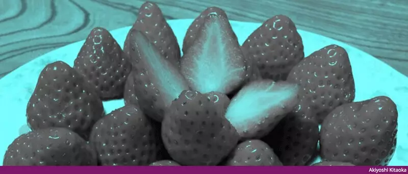
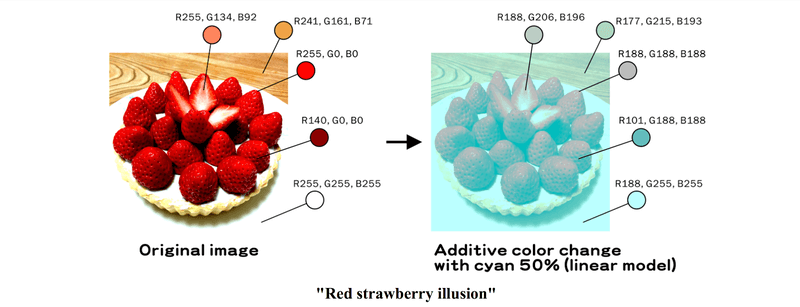
The human brain continuously alters colors to create consistency, referred to as color constancy. The strawberry illusion, where strawberries are seen to have a red color without any red pixels, and the famous dress’ debate (blue-black vs white-gold), prove the point well. The brain creates an image by filling in the blanks about the colors through light and prior knowledge. This is the same way that brain imagery is analyzed, so in essence, your “interpretation” is a constructed version of reality.
Does Brain Color Change in Different States?
There can be slight variations in human brain color depending on how the body operates, but they do not vary significantly from one another. There are some differences in blood flow, oxygen consumption, and other aspects that result in different shades of pink.
If there is any physical activity or even emotional stress, there will be additional blood flow to the brain, because more oxygen needs to be delivered. As a result, the tissue will get a brighter shade compared to normal conditions, and small vessels will become more visible.
During sleep, there are no variations in blood circulation, even if the state of mind changes. The brain is very active at night and maintains the same color as during the day – pink-gray. Oxygen supply and blood flow to the brain are continuous.
Aging results in changes to the tissue itself, which causes a less vivid pink color and more grey. White parts of the brain can turn slightly yellow due to tissue changes.
The occurrence of diseases makes it easier to detect changes in color. For example, inflammation leads to the appearance of reddish regions caused by an increase in blood flow, whereas poor circulation might result in lighter areas of the brain. In addition, some tumors that are denser and have other characteristics that differ from those of the surrounding tissues might be easily noticeable.
An important role in brain color is also played by hydration and the amount of oxygen in the blood. For instance, low oxygen content causes changes in color since there can be a loss of pink hue, making the organ look dull. On top of that, dehydration and exhaustion can impact color somewhat as well.
In general, human brain color remains rather stable in the absence of disease, with only subtle shifts reflecting changes in blood flow and biological activity.
Bottom Line
The color of an active human brain is not gray, as most people believe. It is a combination of two colors that work constantly together to create a unique result. The pink shade appears due to the uninterrupted supply of oxygenated blood in millions of blood vessels in the brain. The gray part, as well as the brain texture, is determined by the structure of brain tissue itself. The proportion of the 40/60 gray-white ratio creates brain coloring and texture.
This is why preserved brains appear to be so different. When someone dies, there is no longer any flow of blood into the tissue nor any access to oxygen. This causes the tissues to turn from pinkish to uniformly gray. The preservation solution used, such as formaldehyde, preserves the brain in this state and may even cause the brain tissue to appear yellow or dim.
So, many people will picture an image of the brain that is a solid gray color. This is based on either what is seen in books or in movies; however, it should be known that this is not what the living brain looks like.
Knowledge about how the brain keeps its colors is also an important element in a larger context of understanding the way that memory and perception operate. Software like memoryOS allows you to learn how the brain actually processes, stores, and retrieves information.
FAQ
Is the human brain pink or gray?
The color of a living brain can be referred to as a mixture of both colors. It is called “brain pink” or “pink-grayish”. Such shades of the brain are created by oxygen-containing blood and its flow via capillaries.
Why is the brain called “gray matter” if it’s pink?
Gray matter is the color attributed to the preserved brain. As soon as the tissue gets stripped of blood, it looks grayish-brown. Therefore, the scientists who studied it first called it gray. However, in a living brain, gray matter is rather pink.
What color is the brain without blood?
If there is no blood in our brain, then the answer to this question is quite simple, as without blood, the brain has a grayish-white color. The dark parts are gray matter, and the light or creamy parts are white matter because of the fatty myelin sheath. This is exactly how brains preserved in museums are.
Can you see your own brain color?
No, it is not possible to observe one’s own brain directly. During surgery, one cannot see one’s own brain as well. Images obtained by means of MRI and CT scans are not in natural colors but rather artificially colored.
Do all animal brains look the same color?
The color of most mammals is the same, although some may differ slightly depending on the amount of gray matter or the proportions of white and gray matter.
Why do brains in movies look so gray?
Most brain shots in films show preserved brains or brain models. Preserved brains look gray as their blood was drained out to keep them from rotting. Filming actual brains would be impossible, as real brains cannot be preserved without losing color.
Contributors